MOUSE
Mouse brain
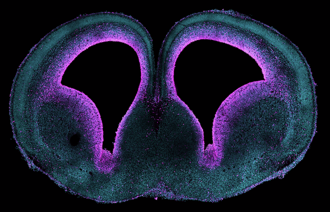
Embryonic mouse brain (E15) showing progenitors in magenta. Nuclei are in blue. Acquired by Nereo Kalebic
MOUSE
Embryonic mouse brain (E15) showing progenitors in magenta. Nuclei are in blue. Acquired by Nereo Kalebic
FERRET
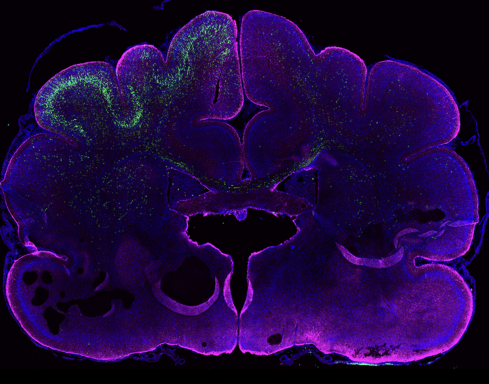
Ferret embryos were electroporated with GFP (green) at embryonic day 37. 20 days later, at postnatal day 16, brain was analyzed and stained by GFAP (magenta). Nuclei are in blue. Acquired by Nereo Kalebic.
.png)
ORGANOIDS
Example of a six-weeks-old human cortical brain organoid stained for progenitor markers SOX2 (green) and Ki67 (magenta) along with DAPI (blue). Acquired by Ilaria Bertani and Stefania Faletti.
EVOLUTION
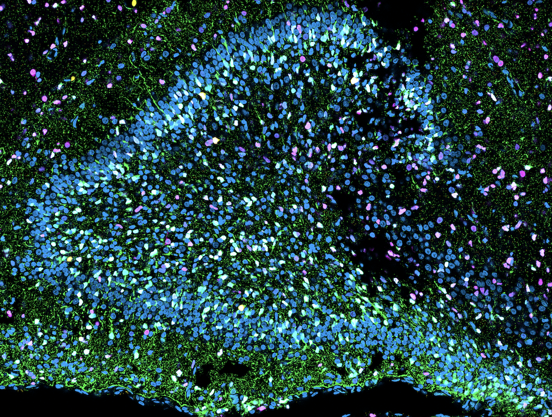
Ferret dentate gyrus (postnatal day 16) stained with HOPX (green) and SOX (magenta). Nuclei are in blue. Acquired by Chiara Ossola
BASAL RADIAL GLIA
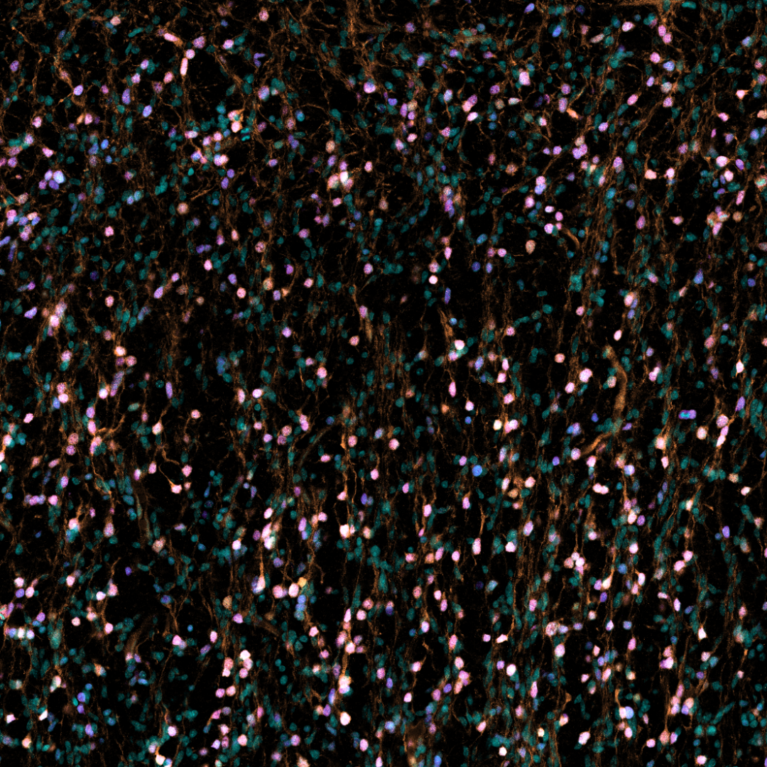
Basal radial glia in the outer subventricular zone of the human fetal cortex (post conception week 14). The progenitors are marked by HOPX (orange) and SOX2 (magenta). Nuclei are in light blue. Acquired by Emanuele Capra.
GLIOBLASTOMA
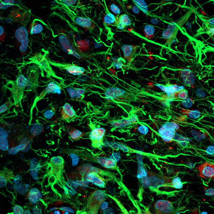
Glioblastoma stem cells in patient-derived glioblastoma organoids. Nestin (green), SOX2 (red); nuclei (blue). Acquired by Carlotta Barelli.
GLIOBLASTOMA
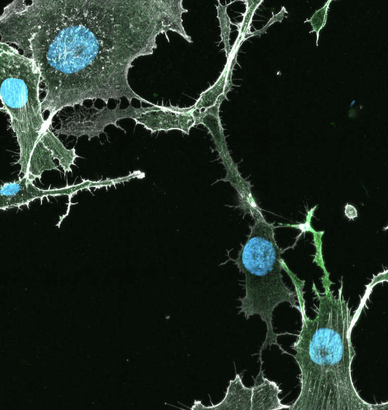
Glioblastoma stem cells growing cell-cell connections that convey resistance to chemotherapy. GFP (green), Phalloidin (white); nuclei (blue). Acquired by Carlotta Barelli.
NEURONS
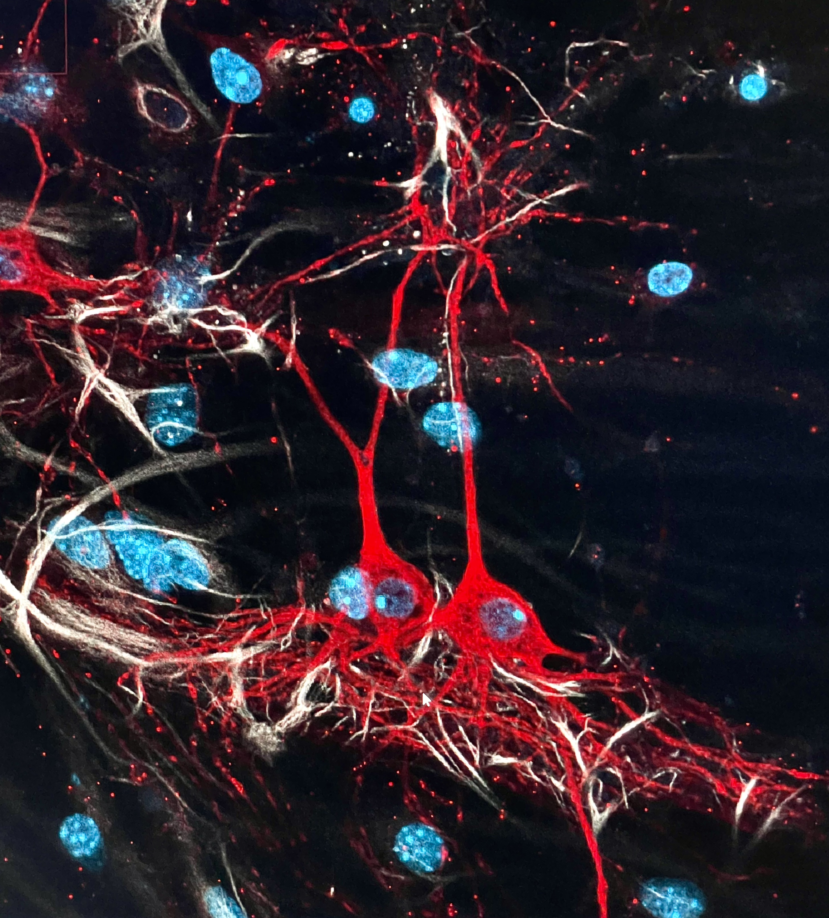
Mouse cortical neurons and astrocytes grown in 2D cell culture. MAP2 (red), GFAP (white), nuclei (cyan). Acquired by Ilaria Bertani.